Health & Performance · Research signal
The Heart Has a Nervous System of Its Own
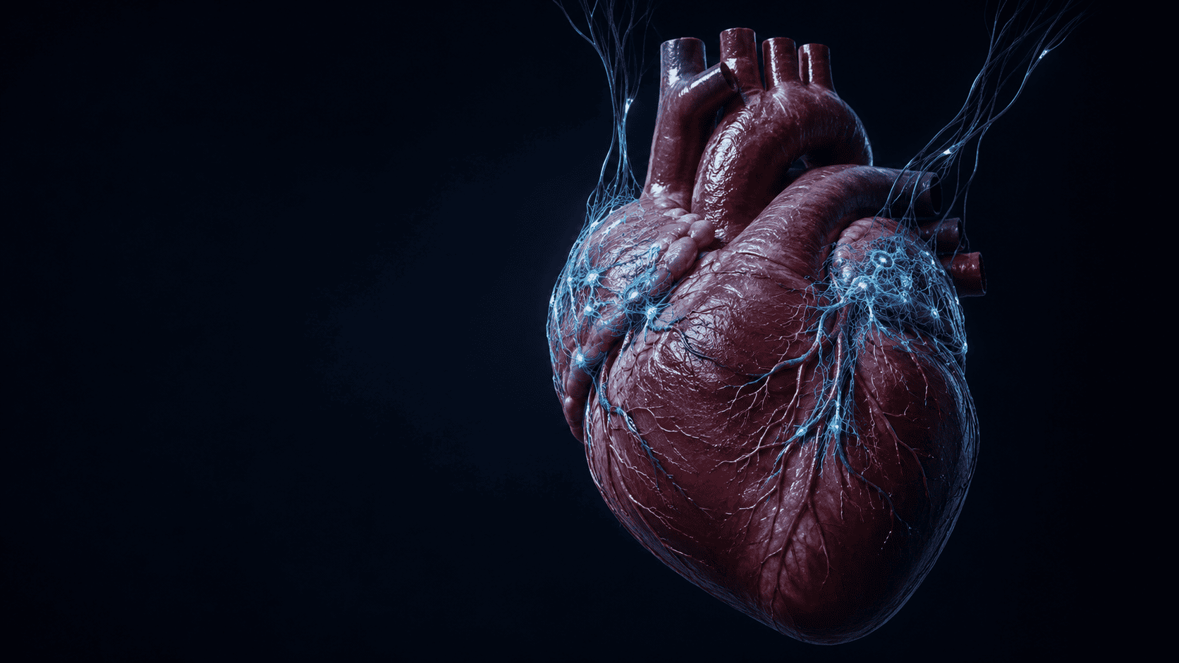
The heart nervous system is a local control layer, not a second mind. Your heart does not think. It does something more biologically precise: local neurons help tune blood flow, rhythm and electrical stability, while different circuits reveal their purpose under different kinds of load.
Humans have intracardiac ganglia, but their cells differ from common animal models.
Npy+ and Ddah1+ populations were activated and removed in controlled experiments.
Multiple neuron types and rhythmogenic firing were mapped without proving consciousness or a human cardiac CPG.
Not a second brain. Not consciousness in your chest. A real, local network of neurons that sits between incoming commands and the heart tissue they are meant to control. New research is beginning to show which cells do what, why some matter most at rest, and why others may become critical only when the system is pushed toward failure.
For most of us, the wiring diagram is simple. The brain decides, an autonomic command travels down a nerve, and the heart obeys. Sympathetic activity speeds it up. The vagus nerve slows it down. The heart is the muscular endpoint.
That diagram is not false. It is missing a layer.
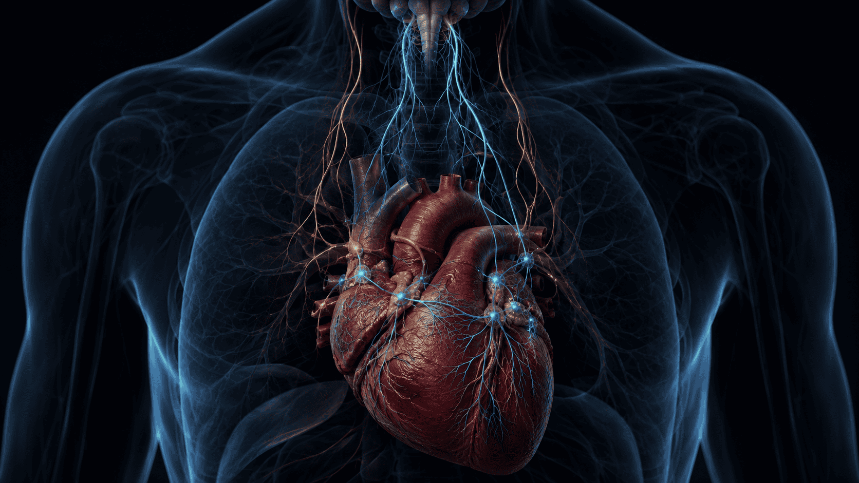
Sitting within and around the heart is a network of neurons called the intrinsic cardiac nervous system, or ICNS. Its cells are clustered in small ganglia, connected to each other, connected to incoming autonomic pathways, and connected to cardiac tissue. They receive information, alter one another’s activity, and modulate the heart’s rate, conduction, contractility, coronary blood flow, and electrical stability.
Some researchers have called this network the heart’s little brain. The phrase is memorable, but it comes with a cost. A brain is not simply any place where neurons exist. The human brain integrates perception, memory, language, planning, self-models, and conscious experience on a scale for which there is no evidence in the heart.
The ICNS is interesting for a different reason. It reveals that physiological control is distributed. The brain is not issuing a separate instruction for every beat. The heart is not acting alone either. Stability emerges from layers of local and central regulation that continuously constrain, correct, and update one another.
In short
The heart contains a local nervous system that is embedded in a larger brain-body network. It does not create the heartbeat in the way the sinoatrial node does, and it does not think in the way the brain does. It modulates how the heart performs.
Research since 2024 has sharpened that picture. Adult zebrafish were found to have multiple types of intracardiac neurons with different molecular identities and firing patterns. A 2025 comparison showed that mouse, pig, and human intracardiac neurons differ substantially, which warns against treating a mouse heart as a small human heart. A 2026 developmental study showed that organs help shape the neurons that innervate them. Then a causal mouse study identified two intrinsic neuron populations with different jobs: one was essential for baseline cardiac function and parasympathetic control, while another protected electrical stability during extreme sympathetic stress.
The result is not a second brain. It is a local control network with a division of labor.
Three systems that are easy to confuse
The strongest version of this story begins by separating three things that popular explanations often merge.
1. The pacemaker
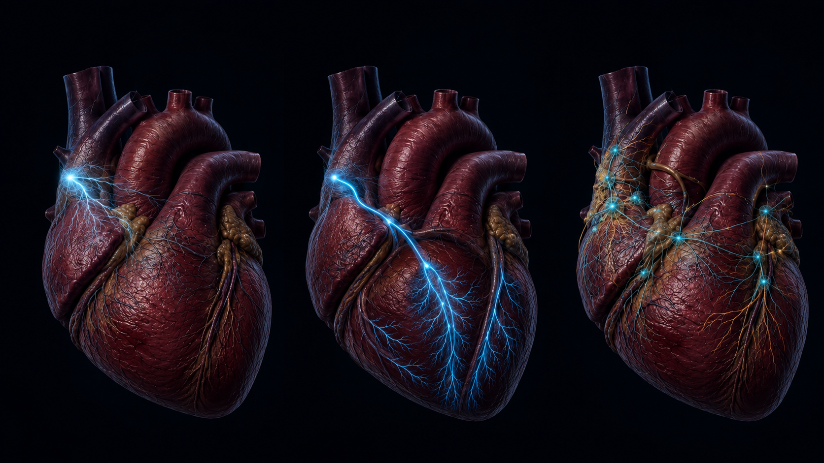
The normal heartbeat begins primarily in the sinoatrial node, a small region of specialized cardiomyocytes in the right atrium. These cells slowly depolarize on their own. When they reach threshold, they fire an action potential that initiates the next beat.
These are not neurons. They are electrically specialized heart muscle cells.
2. The conduction system
The impulse moves through atrial tissue, pauses at the atrioventricular node, and travels through the His-Purkinje network to coordinate ventricular contraction. The history of cardiac physiology was transformed when nineteenth and early twentieth-century anatomists distinguished this specialized conducting tissue from both ordinary muscle and nerves.
3. The intrinsic cardiac nervous system
The ICNS adjusts the performance of the pacemaker and conduction system. It influences when cells fire, how quickly impulses conduct, how forcefully muscle contracts, and how coronary vessels respond. It can also participate in local reflexes and change how incoming sympathetic and vagal activity is translated into a cardiac response.
A simple analogy is useful. The sinoatrial node is the oscillator. The conduction system is the timing distribution network. The ICNS is part of the adaptive control layer. Like all analogies, this one has limits, but it prevents the central error: neurons do not normally replace the sinoatrial node as the source of every beat.
What is actually inside the heart
Intracardiac neurons are concentrated mainly in atrial ganglionated plexuses, often within epicardial fat pads near major vessels and nodal regions. The exact map varies by species. Large mammals and humans do not simply scale up the compact arrangement found in mice.
The network includes several functional classes:
- efferent neurons that influence cardiac tissue;
- afferent or sensory neurons that respond to mechanical and chemical state;
- local circuit neurons that modify activity within the network;
- axons arriving from, or returning toward, extracardiac pathways.
The chemical vocabulary is diverse as well. Acetylcholine is central to parasympathetic cardiac control, while norepinephrine is central to sympathetic effects. Neuropeptides such as neuropeptide Y and vasoactive intestinal peptide add another layer of modulation. A cell’s marker, however, should not be confused with a complete job description. When researchers call a population Npy+ or Ddah1+, they are using gene expression to identify a cell group. The experiment must still show what that population does.
Older diagrams treated intracardiac ganglia as relay stations. Modern work increasingly treats them as a network. The difference is important. A relay passes a message. A network can combine multiple inputs, filter them according to state, and produce different outputs under different conditions.
A global prehistory of the heart-mind idea
Long before anyone could see a neuron, humans noticed that fear, fever, effort, pain, desire, and illness changed the pulse. That observation is nearly universal because the phenomenon is accessible without an instrument. Put a hand on a chest or artery and the body appears to reveal its internal state.

Ancient Egyptian medical texts linked the heart to pulsations felt throughout the body. The Ebers Papyrus describes the physician assessing the heart through vessels leading to body parts. This was not a modern map of circulation, but it was a disciplined recognition that a central organ could be read at a distance.
Mesopotamian diagnostic traditions recorded bodily signs, including changes associated with the heart and pulse, inside systems that mixed empirical observation with divine interpretation. The distinction matters. Ancient medicine often watched the body carefully without separating mechanism, omen, ritual, and prognosis in the way modern research tries to do.
Greek traditions disagreed with one another. Aristotle placed the heart near the center of life, movement, and sensation. The Hippocratic text On the Sacred Disease argued instead that the brain was the source of understanding and emotion. The ancient world did not speak with one heart-centered voice.
In classical Indian medicine, the Charaka Samhita described the heart as a root of major vessels and also as a seat of consciousness. In the Chinese Huangdi Neijing, the heart became the ruling official of an organ-state, associated with the emergence of spirit and clarity. These are sophisticated medical and philosophical models, but translating them directly into modern neuroanatomy would be a category error.
Ibn Sina later described systematic changes in pulse with emotional and physical state. Ibn al-Nafis corrected the route of blood through the lungs. Nahua thought paired face and heart as a way of speaking about formed personhood. Each tradition noticed or organized a different part of the human problem: circulation, pulse, vitality, identity, emotion, or moral character.
None identified the intrinsic cardiac nervous system.
That boundary does not make the old sources irrelevant. It makes them more interesting. They show why the heart became a global metaphor for selfhood. It is rhythmic, audible, responsive to emotion, necessary for life, and partly available to conscious perception. Modern neurocardiology explains some of that responsiveness. It does not prove that every heart-centered cosmology was secretly describing neurons.
The experimental path began much later. Galen observed that hearts could continue pulsing after nerve interruption, even while his circulation model remained wrong. William Harvey established the heart as the driver of a circulating blood system in 1628. Robert Remak described cardiac ganglion cells in 1839. The Weber brothers showed that vagus stimulation could slow or stop the heart. The cardiac conduction system was then anatomically separated from nerves and ordinary myocardium.
In 1921, Otto Loewi supplied one of physiology’s most elegant experiments. He stimulated the vagus nerve of a perfused frog heart, slowing it. Fluid transferred from that preparation then slowed a second heart. The signal could travel in fluid. Neural control of the heart was chemical, not only electrical. The substance he called Vagusstoff was later identified as acetylcholine.
By the late twentieth century, researchers including J. Andrew Armour and Jeffrey Ardell were building a modern model of neurocardiology. Intracardiac neurons were not merely terminal switches. They formed a distributed network with sensory, motor, and local circuit properties. The little brain metaphor emerged from that work. The science was a network claim. The public story often turned it into a consciousness claim.
What the 2024 zebrafish study found
In December 2024, a team led by Konstantinos Ampatzis published a detailed analysis of the adult zebrafish intracardiac nervous system in Nature Communications. The researchers combined single-cell RNA sequencing, anatomical mapping, and direct electrophysiology.
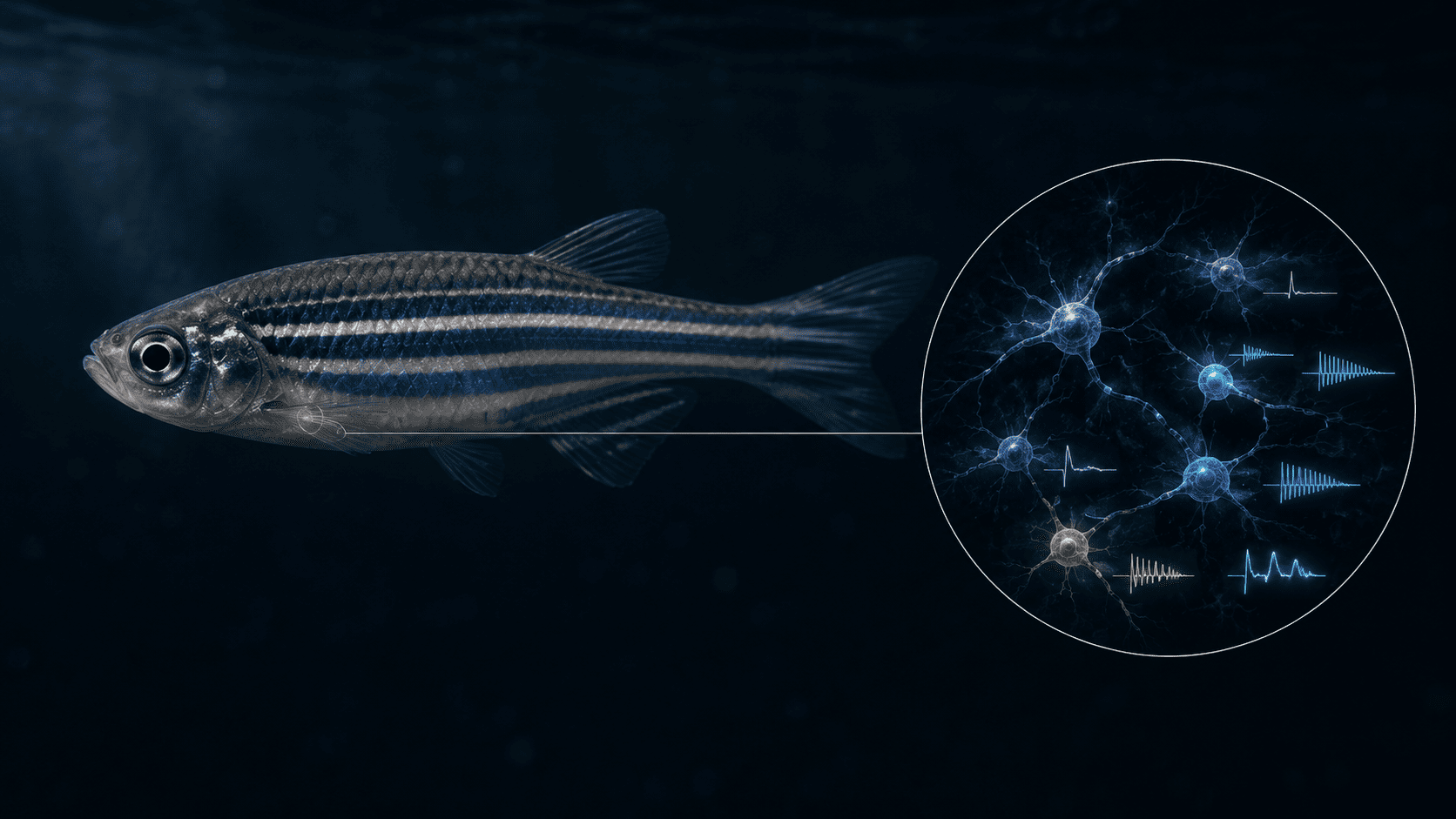
The sequencing dataset contained 9,508 heart cells divided into 22 clusters and eight broad populations. Neuronal cells were a small minority, less than 1.5 percent of the sampled cells. Yet their organization was far from uniform. Most of the mapped neurons were concentrated near valve and sinoatrial regions. Their molecular signatures included cholinergic, catecholaminergic, glutamatergic, GABAergic, and serotonergic features.
When the team recorded individual neurons, it found several firing styles: single-spike, adaptive, repetitive, and bursting. The bursting population was especially provocative because it showed spontaneous rhythmic behavior resembling features found in central pattern-generating circuits.
Central pattern generators are neural networks that help organize repetitive actions such as walking, swimming, or breathing. Their rhythms can be generated or sustained without a conscious command for every cycle. Seeing rhythmogenic properties in intracardiac neurons therefore suggests that the local network may contain more temporal organization than a simple relay requires.
But suggests is the correct word.
The study did not establish that the zebrafish ICNS is a complete central pattern generator. It did not show that neurons normally replace pacemaker cardiomyocytes. In an ex-vivo manipulation designed to provoke synaptic release, neuronal activity altered cardiomyocyte firing and reduced heart rate, but it did not abolish the beat. The authors themselves framed a CPG-like role as a possibility requiring more evidence.
The durable finding is heterogeneity. A small number of neurons can still form a structured, functionally diverse control layer.
The human translation gap
Animal models are indispensable because researchers can record, label, activate, silence, or ablate specific cells in ways that are not ethically possible in living people. The cost is translation.
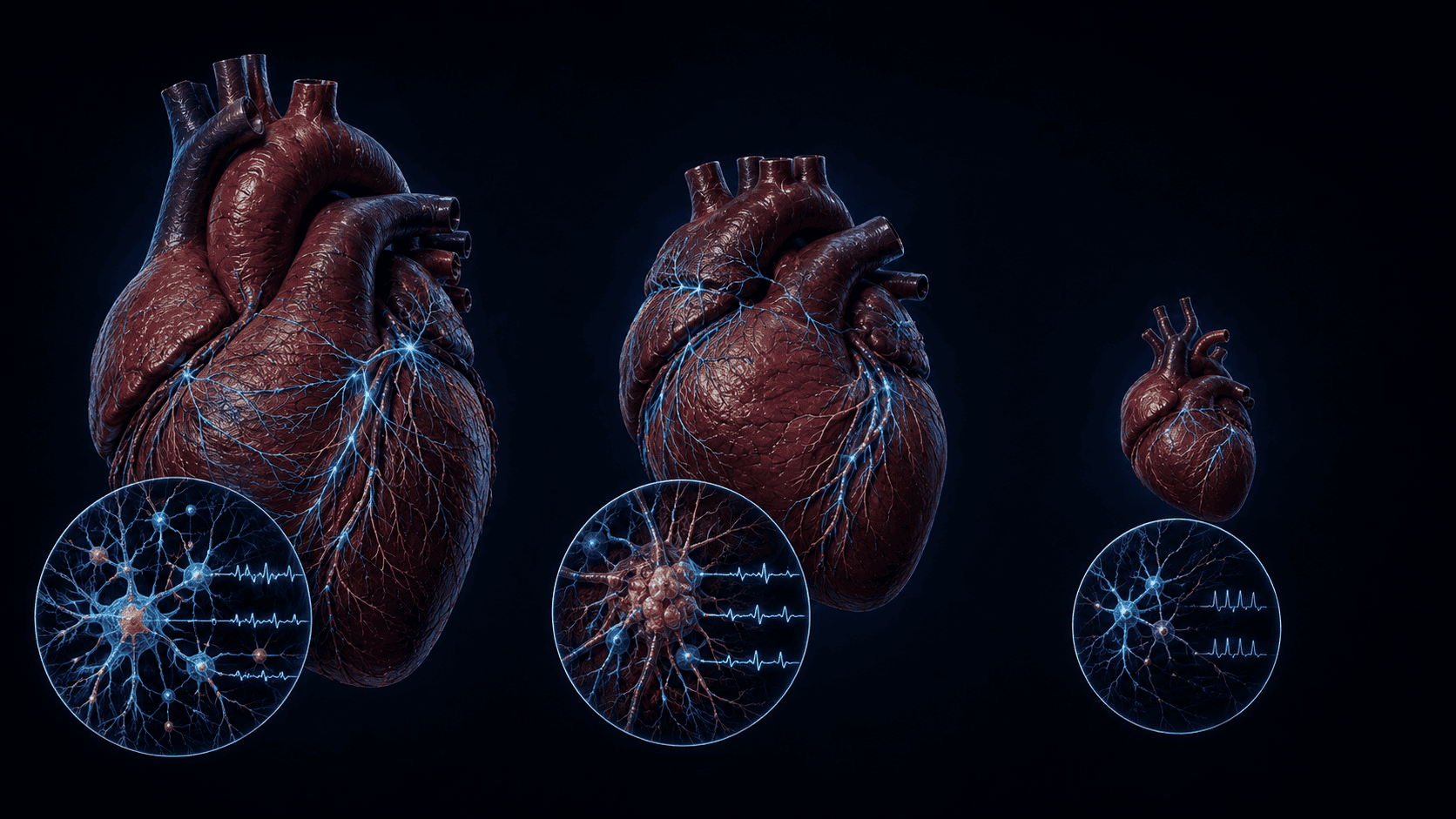
A 2025 study compared intracardiac neurons from donated human hearts with neurons from pigs and mice. It found that the three species are not equivalent control systems at different scales.
Mouse intrinsic neurons were smaller, densely packed, and often received powerful axosomatic inputs. Pig neurons had extensive dendritic trees and integrated more subthreshold cholinergic signals. Human neurons combined properties seen separately in the other species. About 95 percent fired tonically in the tested conditions, and human tissue showed abundant vasoactive intestinal peptide features that did not map neatly onto the mouse or pig pattern.
Neuropeptide Y-positive neurons were identified across species. That does not mean the Npy+ population has an identical role in every species. A shared molecular label is evidence of resemblance, not proof of conserved circuit function.
This matters enormously for the 2026 mouse findings. The mouse study can demonstrate that a targeted population is necessary in a mouse. It cannot by itself establish the same therapeutic target, safe stimulation pattern, or failure mode in a human heart.
The translation gap is not a footnote. It is part of the result.
How the heart helps build its own nervous system
One of the most unexpected advances arrived in a separate Nature paper in May 2026. Researchers traced the development of intrinsic neurons across mouse organs, including heart, pancreas, intestine, and lung.
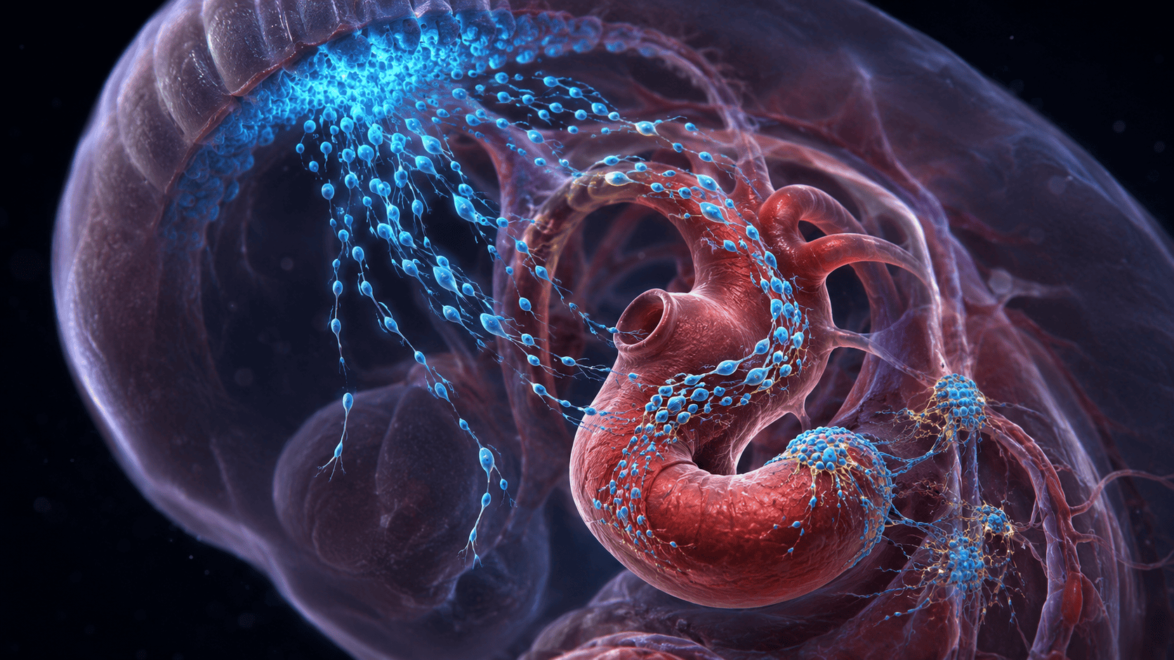
The standard developmental story begins with neural crest cells. These embryonic cells migrate through the body and give rise to much of the peripheral nervous system. The new work found a dual logic. Migratory lineage helped establish a spatial framework, but the local organ environment helped instruct what the arriving neurons became.
In the heart, extracellular matrix and integrin-related signals supported neurogenesis and helped stabilize the stereotyped arrangement of cardiac ganglia. The implication is subtle and important: an organ is not merely a passive destination waiting for generic nerves to arrive. Its tissue environment helps build the nervous system that will later regulate it.
That offers a new way to think about disease and repair. If organ-derived signals shape neural identity during development, changes in extracellular matrix, inflammation, fibrosis, or tissue composition later in life may also influence neural maintenance and remodelling. That extension is a hypothesis, not yet a proven therapeutic mechanism, but it creates a research bridge between developmental biology and adult heart disease.
What the 2026 mouse study demonstrated
Preferential vagal input; parasympathetic heart-rate control and coronary perfusion. Ablation caused severe failure in the mouse model.
Predominantly sympathetic input; protection of electrical stability became critical during extreme experimental stress.
In July 2026, researchers led from Yale School of Medicine published a causal study in Cell titled The intrinsic cardiac nervous system is essential for cardiac function and survival.
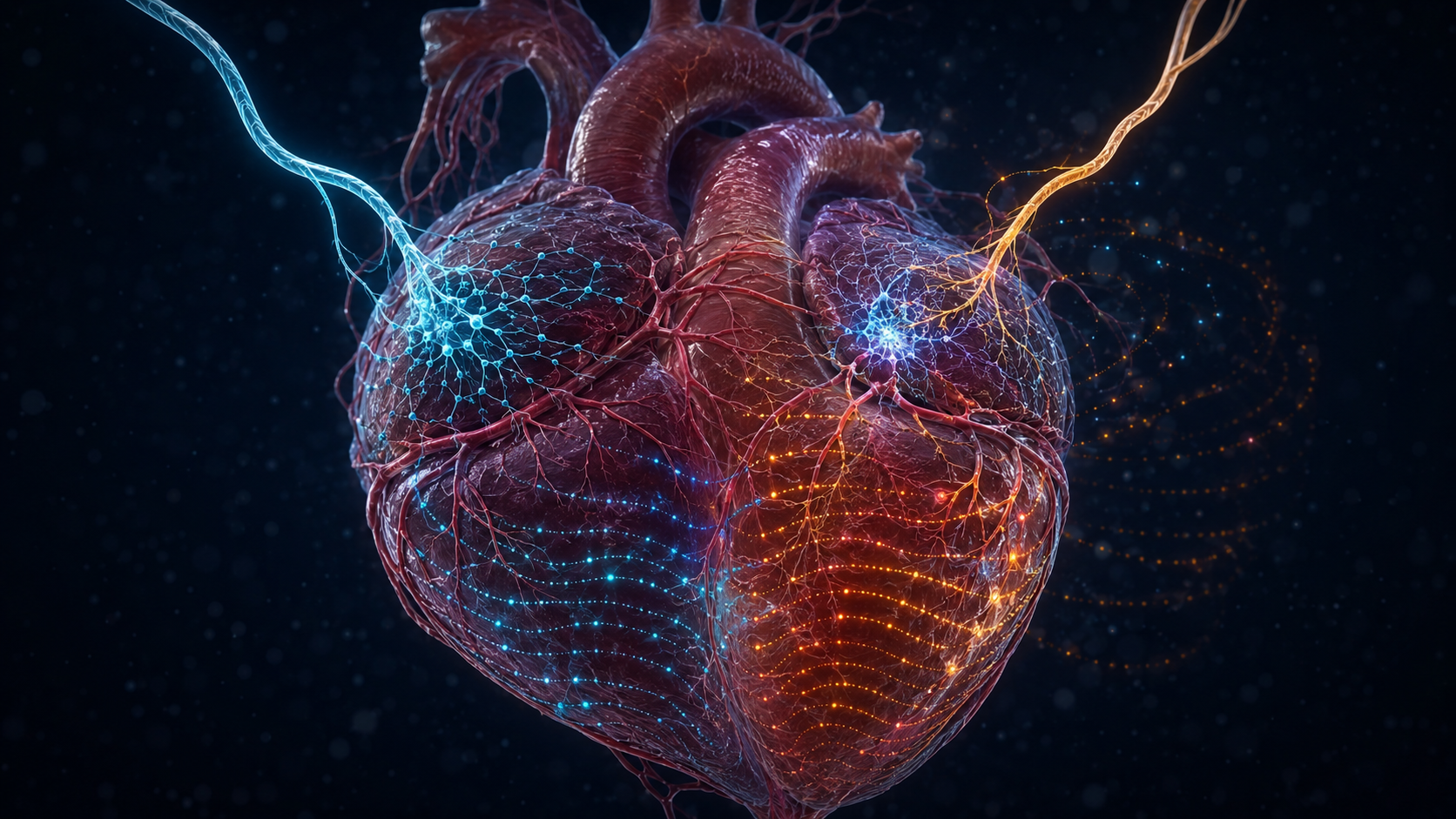
They combined single-cell transcriptomics, genetic labelling, high-resolution imaging, targeted activation, and cell ablation. Two major intrinsic neuron populations stood out, identified by expression of the genes Npy and Ddah1.
Npy+ neurons: baseline control
The Npy+ population received preferential vagal input and was central to parasympathetic control. Activating these neurons slowed heart rate. Removing the population abolished normal parasympathetic heart-rate control and disrupted coronary perfusion and baseline cardiac performance so severely that the heart could not sustain function.
This is what load-bearing means in a biological network. A population can be numerically tiny and still occupy a critical control point.
The marker should still be read carefully. The result does not mean that the NPY molecule alone is the master switch of cardiac survival. It means that the cell population identified by Npy expression had that causal role under the experimental conditions.
Ddah1+ neurons: protection under extreme stress
The Ddah1+ population received predominantly sympathetic input and behaved differently. Under baseline conditions, its loss did not produce the same immediate collapse. Its importance emerged when sympathetic demand became extreme.
During stress paradigms, including pharmacologically induced sympathetic overactivation, activating the population increased resilience. Disrupting it made the heart more vulnerable to electrical instability and sudden death.
This is a different kind of essentiality. Some components keep a system operating every day. Others become essential only near the boundary of failure.
That division of labor has wider implications. A calm laboratory measurement can miss the purpose of a circuit designed for rare extremes. Biological function often appears only when the system is challenged.
The study therefore changes the question. Instead of asking whether the heart has local neurons, researchers can ask which cells stabilize which variables, under which conditions, through which inputs, and at what cost.
A transplanted heart as a natural experiment
Heart transplantation is often used as proof that the heart’s little brain can run the organ without the brain. That is not quite the lesson.
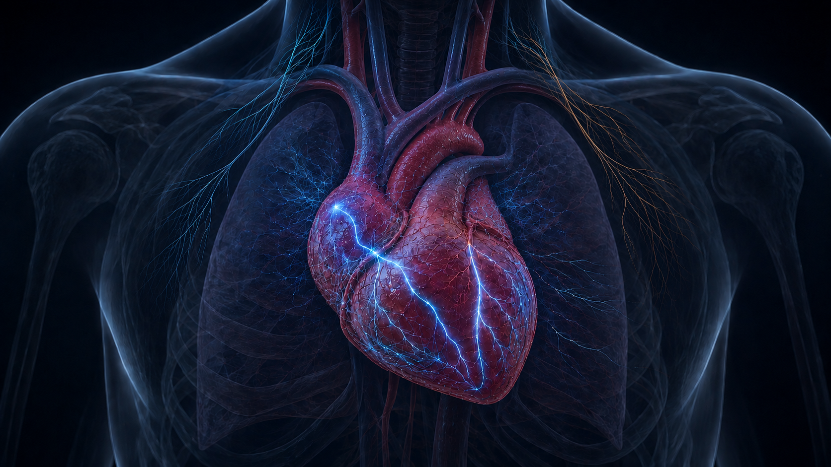
During transplantation, the donor heart’s extrinsic sympathetic and parasympathetic connections are severed. The transplanted heart initially loses normal moment-to-moment neural control from the recipient. Resting heart rate is often higher. Exercise responses and reflex changes can be blunted or delayed. Heart-rate variability is reduced early after transplant. Some reinnervation may occur over time, but it is variable, incomplete, and often more evident for sympathetic than parasympathetic pathways.
The heart still beats because the sinoatrial node is myogenic. Its pacemaker cells generate spontaneous electrical activity without requiring a new vagal or sympathetic command for each beat.
Parts of the donor heart’s intrinsic neural architecture may remain with the organ and continue local interactions, but they do not replace the full lost connection to the recipient nervous system. Transplantation therefore demonstrates two things at once:
- beat generation is intrinsic to cardiac pacemaker tissue;
- normal adaptation depends on wider neural integration.
Autonomy and dependence are not opposites here. The heart possesses both.
When heart damage becomes network damage
A myocardial infarction is usually described as muscle injury followed by scar. That description is correct and incomplete.
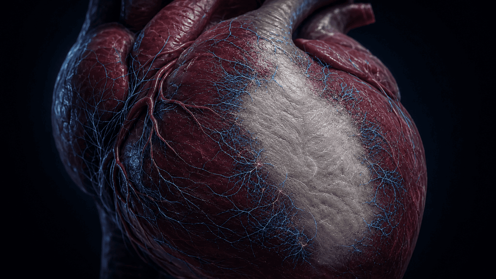
The cardiac nervous system also remodels.
In a porcine model of healed myocardial infarction, researchers found structural and functional changes in intrinsic cardiac neurons. Sensory responses from infarcted tissue were attenuated, while signals from border and remote zones were differently preserved. Local circuit processing changed and network connectivity was reduced. The researchers described a neural sensory border zone around the damaged area.
Extrinsic sympathetic nerves can remodel and sprout after injury as well. Uneven innervation, fibrosis, changed receptor expression, and altered electrical tissue create a potentially arrhythmogenic combination.
This produces an important systems insight: after heart damage, the controller and the controlled tissue can change together. An arrhythmia may not be explained by scar alone or nerve activity alone. It can emerge from their interaction.
The same principle may matter in heart failure, diabetes, chronic inflammation, and ageing. Disease changes the organ that neurons regulate. It can also change the neurons, synapses, receptors, and feedback signals doing the regulating.
Why HRV is an output, not a window into a heart brain
Heart-rate variability, or HRV, measures variation in the time between heartbeats. It is valuable in research and can be useful in longitudinal self-tracking. It is also one of the easiest metrics to overinterpret.
Short-term HRV is influenced by vagal modulation, breathing rate and depth, resting heart rate, posture, age, physical activity, time of day, sleep, alcohol, caffeine, meals, medication, ectopic beats, recording length, and the device or algorithm used. ECG and optical wrist measurements do not always yield interchangeable data.
Respiratory sinus arrhythmia, the rise and fall of heart rate across the breathing cycle, is often used as a proxy for cardiac vagal activity. It is not a fixed, direct readout of central vagal tone. Breathing mechanics and the transfer characteristics of the cardiovascular system matter.
Slow breathing can increase certain HRV measures during the session and affect baroreflex and cardiorespiratory coupling. That does not prove that a person has strengthened a specific ICNS circuit. Nor does one low morning score prove failed recovery.
The more accurate interpretation is this:
HRV is an output of a coupled system, not a direct dashboard of one hidden controller.
For Health & Performance, that makes context more important, not less. The useful question is not whether a number is good in isolation. It is whether repeated measurements, taken under comparable conditions, move with sleep, load, symptoms, training, stress, illness, and recovery.
Why emotion really can be felt in the chest
Rejecting heart consciousness does not mean chest-based emotion is imaginary.
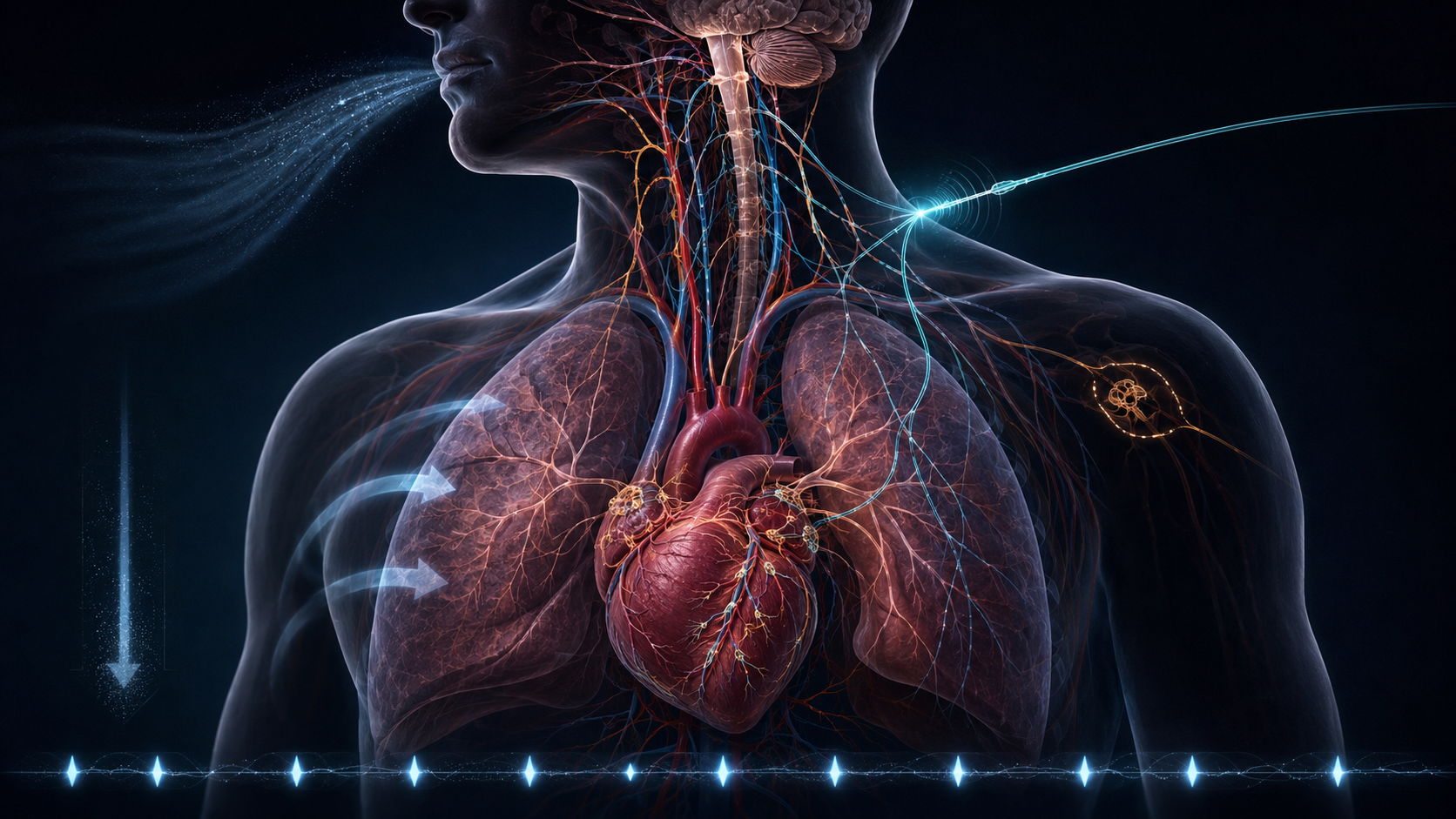
Emotion is a whole-body process. Threat can change breathing, vascular resistance, catecholamine release, muscle tension, and cardiac timing within seconds. Sensory pathways carry information from the heart and blood vessels toward the spinal cord and brainstem. Baroreceptors in the aortic arch and carotid sinus detect stretch. Cardiac afferents respond to mechanical and chemical conditions. Brainstem nuclei integrate that information with respiratory and other visceral signals. Higher networks, including insular and limbic regions, contribute to the conscious sense of bodily state.
What you feel in the chest is therefore real interoception. It is the brain’s experience of signals arising from a changing body, combined with expectation, context, memory, and attention.
The causal arrows run both ways. A frightening interpretation can intensify cardiac sensation. A sudden cardiac change can alter attention and emotion. Breathing can modify both physiology and perception. None of this requires autobiographical memory to be stored in the heart.
The heart is part of emotional experience without being a second seat of consciousness.
The treatment paradox
If local cardiac neurons help cause or sustain arrhythmia, destroying a ganglionated plexus can look attractive. If they protect the heart and maintain normal control, stimulating or preserving them can look equally attractive.
Both intuitions can be correct in different circuits and wrong when applied indiscriminately.
Ganglionated plexus ablation has been studied in atrial fibrillation and cardioneuroablation is being explored for selected reflex syncope and vagally mediated bradyarrhythmias. Results have been mixed. In the AFACT surgical atrial fibrillation trial, adding ganglionated plexus ablation did not produce the expected recurrence benefit and increased major adverse events. Other studies have likewise shown that broad plexus ablation is not automatically superior to established approaches.
The new cell-type evidence offers a possible explanation. A ganglion is not one function. It can contain neurons with different inputs, transmitters, targets, and roles. A coarse intervention may remove a pathological influence, a compensatory influence, and an essential protective influence at the same time.
Future neuromodulation may become more selective by location, cell identity, firing pattern, or physiological state. That is a research direction, not a current promise. The closer science gets to specific circuits, the less defensible it becomes to speak of stimulating or ablating the heart’s nervous system as if it were one switch.
What this does not mean
The evidence supports the following:
- the heart contains real neurons organized into local ganglia and networks;
- these neurons are molecularly and electrophysiologically diverse;
- some local circuits can integrate signals rather than merely relay them;
- targeted neuron populations have causally distinct roles in mouse heart function;
- communication between cardiovascular system and brain is bidirectional;
- human intracardiac neurons exist and differ in important ways from common animal models;
- injury and disease can remodel neural control as well as muscle.
The evidence does not support the following:
- that the heart is conscious;
- that it stores autobiographical memories;
- that love, intuition, or moral wisdom is generated by intracardial ganglia;
- that a wearable can directly measure the health of the ICNS;
- that the mouse Npy+ and Ddah1+ division has already been proven in humans;
- that vagus stimulation, breathing exercises, or ganglion ablation are universally beneficial;
- that ancient heart metaphors were descriptions of modern neurocardiology.
Precision is not a way of making the story smaller. It reveals what is genuinely new.
The bigger pattern
The ICNS belongs to a wider biological principle: control in the body is distributed.
The gut has an extensive enteric nervous system. Spinal circuits execute fast reflexes without waiting for conscious deliberation. The pancreas integrates local metabolic and neural signals. Immune and endocrine networks coordinate through feedback, thresholds, delays, and tissue-specific messages. The brain remains unmatched in cognition and integrated behavior, but it does not micromanage every physiological event.
Distributed control creates resilience. Local systems can respond quickly and preserve function when central input is delayed or disrupted. It also creates complexity. A local response that helps in one context can become harmful in another. A protective stress circuit may be silent at rest. A disease can change both the tissue and the controller trying to stabilize it.
That is why training, recovery, and resilience cannot be reduced to one metric. Breathing, sleep, posture, blood volume, temperature, inflammation, medication, fitness, expectation, and emotional state interact through overlapping loops. A higher HRV value, a slower resting pulse, or a calmer feeling may be informative. None is the system by itself.
The heart is not a passive pump waiting for instructions. It is also not a mind in the chest. It is an electrically self-starting, mechanically powerful, chemically active, neurally regulated organ embedded in a continuous brain-body conversation.
The most interesting finding is not that the heart has a second brain. It is that life depends on intelligence-like organization at many levels, without every level needing consciousness.
Why this matters here
This Health & Performance hub is not about chasing one perfect number. It is about understanding how coupled systems adapt under load.
Heart-rate variability, breathing pattern, stress response, perfusion, rhythm stability, and recovery capacity are not isolated outputs. They emerge from layered regulation. Some of that regulation is central. Some is local. Some is mechanical, chemical, or neural. Much of it changes with context.
That changes what training resilience means. You are not sending commands to a passive machine. You are exposing a distributed biological system to signals, loads, and recovery conditions from which it adapts. The practical lesson is not to worship the heart’s little brain. It is to respect the whole control architecture.
Research sources
Sources and evidence trail
Core current research
- Qian J. Xu et al. (2026), “The intrinsic cardiac nervous system is essential for cardiac function and survival,” Cell. Full text and DOI
- Yale School of Medicine (2026), “‘Little Brain on the Heart’ Is Essential for Cardiac Function.” Research summary
- Hsu et al. (2026), “Developmental logic of organ-specific intrinsic neurons,” Nature 655, 429-437. Full text and DOI
- Pedroni et al. (2024), “Decoding the molecular, cellular, and functional heterogeneity of zebrafish intracardiac nervous system,” Nature Communications 15, 10483. Full text and DOI
- Tompkins et al. (2025), “Comparative specialization of intrinsic cardiac neurons in humans, mice and pigs,” The Journal of Physiology. Full article and DOI
Anatomy, physiology and translation
- Fedele and Brand (2020), “The Intrinsic Cardiac Nervous System and Its Role in Cardiac Pacemaking and Conduction.” Open-access review
- Giannino et al. (2024), review of the intrinsic cardiac nervous system. Open-access article
- Hanna et al. (2021), “Innervation and Neuronal Control of the Mammalian Sinoatrial Node.” Circulation Research
- “Early Denervation and Later Reinnervation of the Heart Following Cardiac Transplantation” (2016). Open-access review
- Rajendran et al. (2016), intrinsic cardiac neural remodelling after myocardial infarction. The Journal of Physiology
Measurement and clinical caution
- Hayano and Yuda (2019), “Pitfalls of assessment of autonomic function by heart rate variability.” Open-access review
- Grossman (2024), review of respiratory sinus arrhythmia and cardiac vagal tone. PubMed
- Laborde et al. (2022), meta-analysis of slow breathing and psychophysiological outcomes. PubMed
- AFACT trial (2016), ganglionated plexus ablation in surgical atrial fibrillation treatment. PubMed
Historical source trail
- Papyrus Ebers, transliteration and translation project. Science in Ancient Egypt, Leipzig
- Hippocrates, On the Sacred Disease. English translation
- Aristotle, Parts of Animals. English translation
- Charaka Samhita, passages on heart and vessels. English translation
- Huangdi Neijing, “Discourse on the Secret Treatise of the Spiritual Orchid.” Chinese Text Project
- Ibn Sina, Canon of Medicine. Digitized English text
- Ibn al-Nafis and the pulmonary circulation. Historical review
- Florentine Codex. Digital manuscript
- William Harvey, De Motu Cordis. Digitized edition
- Remak biography and 1839 cardiac ganglion description. Historical review
- Otto Loewi and the Vagusstoff experiment. Historical review
- J. Andrew Armour (2008), “Potential clinical relevance of the little brain on the mammalian heart.” Experimental Physiology
Can you separate the layers?
Choose one answer per question. The options are shuffled whenever you try again.
Which cells normally initiate the heartbeat?
In which species were Npy+ and Ddah1+ functions tested causally?
Why is HRV not a direct measurement of the ICNS?
Answer all three questions to see your result.